
Behind the Scenes - Inside our New Lab for Blood & Urine Diagnostics
When your pet has blood or urine taken during a consultation, that sample is taken straight into our in-house laboratory. Having a well-equipped lab on site allows us to begin testing most samples immediately, rather than always sending samples away and waiting days for results.
A Closer Look at Our Blood Testing Process
The first step is deciding which type of blood profile best suits your pet and the reason for testing. Once the test profile is selected, the blood sample is prepared for analysis and run through two key machines in our lab
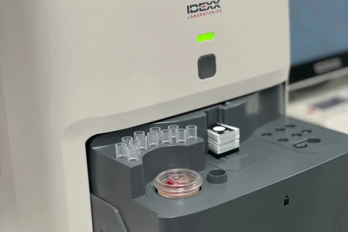
The Catalyst One Laboratory machine is used for biochemistry testing. This means it measures enzymes and substances in the blood that tell us how organs such as the liver and kidneys are functioning.
The blood sample is placed into the machine along with specialised testing slides.
Inside the machine, the blood is spun so the red blood cells separate from the serum.
The serum is then analysed for specific enzymes and markers.
While the Catalyst One focuses on biochemistry, the ProCyte machine handles haematology. This means it looks at the cells in the blood.
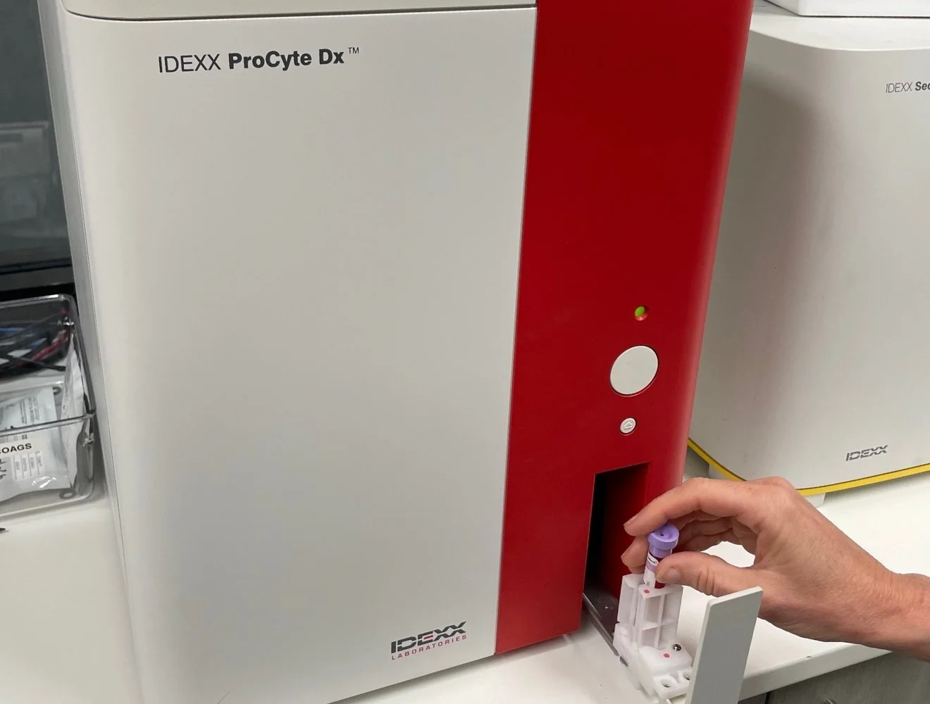
The ProCyte Laboratory machine analyses red blood cells, white blood cells, and platelets.
It uses advanced technology including laser analysis and cell flow measurement to identify and count different cell types.
This allows us to see things like inflammation, infection, anaemia, or how your pets body is responding to illness.
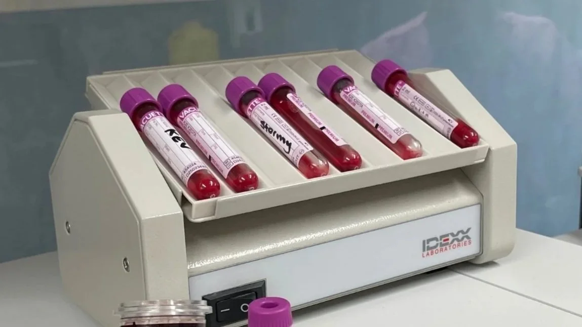
Keeping samples accurate
Blood samples are gently mixed while other testing is underway.
This prevents cells from settling and ensures the results we receive are as accurate as possible.
Accuracy is essential, especially when results help guide diagnosis or treatment decisions.
The importance of vet checks with a microscope and blood smear
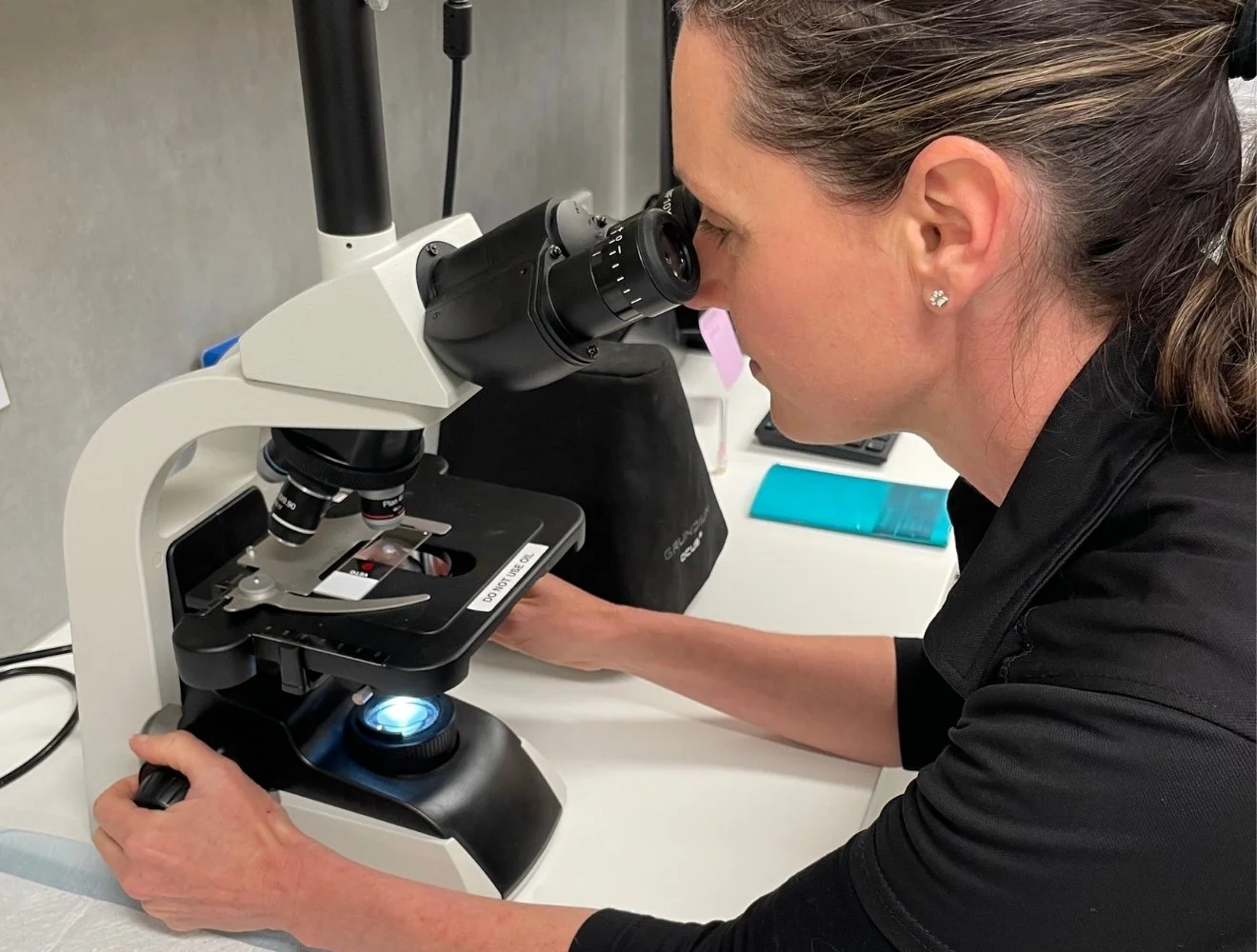
Even with advanced laboratory technology, there are times when nothing replaces the trained eye of a veterinarian looking directly at a sample under the microscope.
A blood smear involves spreading a thin layer of the blood sample onto a glass slide so individual cells can be examined under a microscope. It allows our vets to visually assess cell shape, size, and distribution, and double check automated results.
Blood smears are particularly helpful for confirming platelet counts and identifying subtle changes that machines may not fully capture.
This approach blends technology with hands on clinical judgement, helping us make well informed decisions.
Looking even closer with digital microscopy and remote review
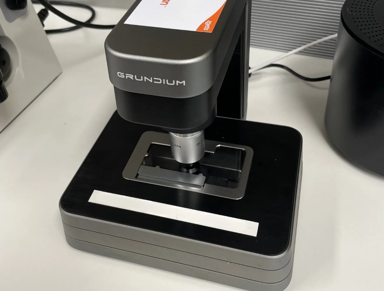
Alongside our in house blood diagnostics machines and microscope work, we often use a digital microscope system called Grundium for blood and urine samples.
This technology allows us to capture high quality images of blood smears and other samples directly through the microscope.
Instead of relying only on what we can see in the moment, we are able to take clear images of individual cells, feathered edges, and areas of interest on the slide.
The main advantage of the Grundium system is that it allows us to securely share these images with external pathology laboratories or specialist teams for review.
If a sample shows changes that would benefit from a second opinion, or if we want confirmation of subtle findings, we can send images instantly rather than physically sending slides away.
This means
Faster specialist input
Less delay in decision making
No risk of slides being damaged in transit
Clear visual records attached to your pet’s case
Once we have finished processing blood samples, either in house or with additional support (when needed) we then move on to the next part of the lab process, urine testing, which provides another important piece of the overall health picture.
A Closer Look at Our Urine Testing Process
Urine testing is often performed alongside blood testing because it provides important information that blood tests alone cannot show. Together, these results help us understand how well organs such as the kidneys are functioning and can highlight issues within the urinary system early.
Urine testing is particularly useful when we are investigating changes in drinking or urination habits, urinary tract infections, bladder concerns, kidney disease, diabetes, or more subtle health changes that may not yet be obvious.
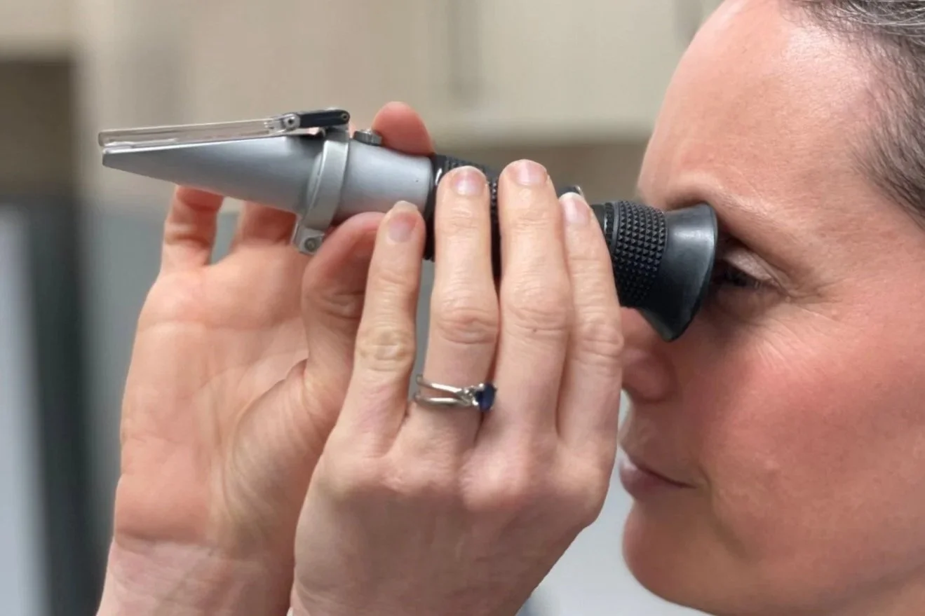
One of the first steps in urine testing is measuring urine concentration, also known as specific gravity.
This tells us how concentrated or diluted the urine is and helps us assess;
How well the kidneys are doing their job
Whether a pet may be drinking too much or too little
How the urine results relate to blood test findings
This step provides important context before we look at anything else in the sample.
Screening with a urine dipstick
We also use a urine dipstick as an initial screening tool.
Dipsticks can give us information about glucose levels, blood in the urine, urine pH, ketones which are particularly important for diabetic patients and other markers that may indicate inflammation or infection. While dipsticks are helpful, they are only one part of the overall assessment.
Analysing urine with IDEXX SediVue DX
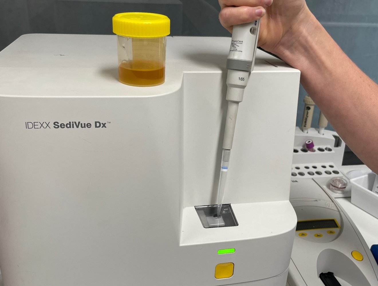
The urine sample is also analysed using our IDEXX SediVue DX machine.
This technology examines urine sediment and helps identify and count
Red blood cells
White blood cells
Crystals
Bacteria
Other microscopic material
The SediVue DX also captures clear images of the sample, which allows us to review findings carefully and explain results when needed. It supports fast and consistent analysis and can be especially helpful for identifying urinary tract infections, crystal formation, and early changes that may affect bladder or kidney health.
While automated urine analysers are excellent tools, they do not replace the value of an experienced eye. Combining technology with hands on microscopic examination gives us the most accurate and reliable results.
Using a centrifuge for closer examination
Even with advanced technology, we also routinely use a centrifuge as part of our urine testing process.
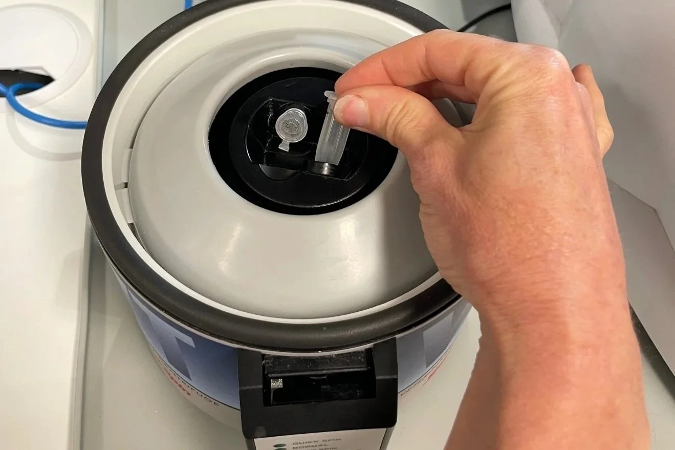
We use a StatSpin veterinary centrifuge to spin the urine sample. This process allows heavier particles in the urine to settle at the bottom of the tube, concentrating the sediment for examination under a microscope.
Centrifuging the urine makes it easier to
Examine cells and crystals under the microscope
Confirm automated results
Detect subtle changes that machines may not fully capture
By combining a layered approach of urine concentration testing, dipstick screening, automated analysis with the SediVue DX Centrifuging and microscopic examination, we gain a thorough understanding of what is happening within your pet’s urinary system.
What happens once the results are ready
Once blood and urine testing is complete, the results are brought together into a diagnostics report. This is where technology steps back and clinical expertise steps forward.
Interpreting laboratory results is not about looking at numbers in isolation.
Our vets consider the full picture, your pet’s history, age, breed, lifestyle, symptoms, and physical examination findings, alongside the laboratory data. Patterns, trends, and subtle changes are often more important than a single value.
From there, your vet can explain what the results mean, whether further investigation is needed, or whether monitoring over time is the best next step.
When treatment is required, results guide decisions around medication, diet, ongoing testing, or referral, always tailored to your individual pet.
“Laboratory testing gives us vital information, but it is the experience and judgement of the veterinary team that turns those results into a thoughtful care plan focused on your pet’s comfort, health, and quality of life.”